SISCOM analysis
Source code GitHub
Introduction
In Nuclear Medicine, it has been developed a program to localize with high precision the epileptogenic focus. This has been motivated by the need of surgery procedure to remove the origin of crisis in drug-resistant epilepsy. The results from DTI (MRI), video-EEG, SPECT and PET are analysed by a multidisciplinary committee formed by the nuclear doctor, the neuroradiologist, the neuropsychologist, the neurosurgeon, among others. The group of Biomedical Images from the University of Barcelona and the Epilepsy Unit of Hospital Clínic of Barcelona have been using the SISCOM methodology.
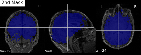
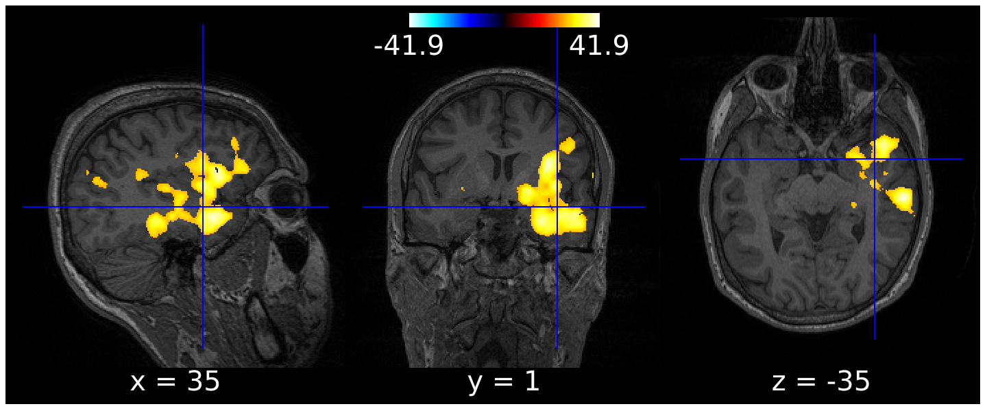
It is known that at the beginning of a crisis (ictal state) the blood flow at the focal region increases compared to interictal or basal state. Using [Tc99m]-HMPAO or [Tc99m]-ECD blood perfusion radiotracers, capable to go through the blood-brain barrier and stay inside, is obtained an instantaneous image of the injection or ictal moment using SPECT after the crisis. After few days with no crisis another SPECT is done to record the interictal state. The subtraction of these images remarks the focus. This information is merged with the RM that will provide us with anatomical information. This procedure is known as SISCOM technique (Subtraction of Ictal SPECT CO-registered with MRI). After the post-processing of the results the evaluation is done with simulated images, Monte Carlo projections, comparing a theoretical focus with the experimental focus.